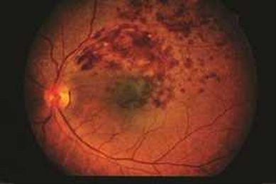
What is a Branch Retinal Vein Occlusion?
What is a branch retinal vein occlusion? As opposed to a central retinal vein occlusion, a branch retinal vein occlusion occurs when some of the smaller veins of the eye, responsible for carrying blood away from the nerve cells of the retina, become blocked. This blood carries oxygen and nutrients, and if the veins become blocked, …
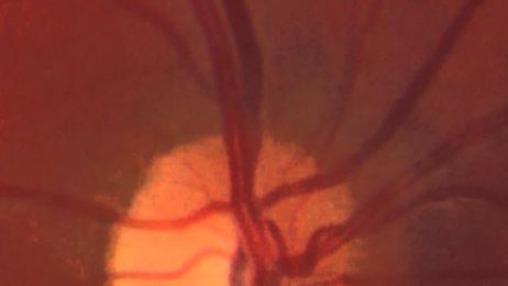
What Is Central Retinal Vein Occlusion (CRVO)?
Central retinal vein occlusion (CRVO) is a blockage of the main vein in the retina.

What's digital retinal photography?
You may have had a retinal photograph taken as part of your eye examination. One purpose of this photograph is to give the optician a clear view of the small...
'Wonderful' Cholesterol Ratio and Retinal Occlusion: Should She Take a Statin?

Retinal Detachment: What Is a Torn or Detached Retina?
A torn retina is when the retina tears in one or more places. A detached retina is when the retina is lifted off the wall of the back of the eye.

How inflammation may play a role in retinal disease
Whenever I deem a case of viral conjunctivitis to be significant enough to warrant the prescription of a topical steroid, I have a very brief discussion with the patient beforehand.

How to Take Retinal Images with a Smartphone
In this video, Dr. Pedro Acevedo describes how to use a smartphone to capture retinal images. This method is particularly helpful in the evaluation of retinopathy of prematurity (ROP) and for use in t

Retinal light exposure after cataract surgery, what are the risks?
Phototoxicity is a current vision health concern and there is evidence that UV and blue-violet light may cause adverse effects to the eye. Blue-violet light sources include the sun, but also the widespread light-emitting diode (LED) technologies, resulting in around-the-clock exposure. Chronic exposure to blue-violet light, among other factors, may contribute to retinal diseases such as age-related macular degeneration (AMD), or speed-up AMD progression after cataract…

What Causes Retinal Detachment?
This articles tackles the topic of retinal detachment and how regular visits to an eye doctor may help prevent it from happening.

What Are the Benefits of a Retinal Screening?
A retinal screening allows your eye doctor a wider view of your eye where they can assess for various health conditions such as diabetes.

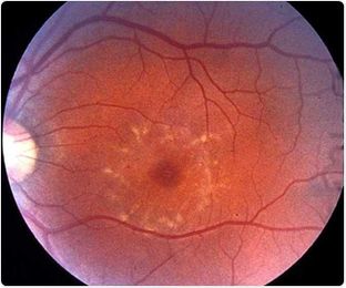
What is Fundus Flavimaculatus?
Fundus Flavimaculatus is a genetic condition which is considered to represent one of two ends of the spectrum of a disease which is comprised of the presentation of retinal flecks. Research has shown that the ABCA4 and PRPH2 genes may be linked to the onset of the condition, as well as the overproduction of vitamin A.
Why you should NOT ignore new floaters in your vision
Sudden flashes or floaters (especially when noticed in just one eye) could<br>be the first warning signs of a retinal detachment or retinal tear. Do not<br>let new flashes/floaters just linger, hoping they will go away. If your<br>symptoms are actually due to a retinal detachment, PROMPT diagnosis and

Retinal Degeneration Can Be Prevented By Exercise
Our eyes are the windows to our souls. Indeed, our eyes have many functions, including light reception, light perception and vision. Without our eyes we cannot perceive our environment, the

Side Effect Of Retinal Detachment May Be Prevented By Ranibizumab
Proliferative vitreoretinopathy (PVR), or the formation of scar tissue in the eye, is a serious, sight-threatening complication in people recovering from surgical repair of retinal detachment.

What are Aberrations of the Eye?
The human eye can be affected by a number of aberrations which can reduce the quality of retinal images and general visual perception. Research has shown that age increases the probability of developing higher order aberrations in particular.

Retinal Implants Could Restore Sight to More Than 2 Million People
If the implants are successful in human trials, they could help restore vision for 2 million people with age-related macular degeneration.

Retinal-scan analysis can predict advance of macular degeneration, study finds
A new computer algorithm could help physicians predict whether a patient’s macular degeneration will progress within a year’s time to the "wet" stage.

Retinal changes may serve as measures of brain pathology in schizophrenia
Schizophrenia is associated with structural and functional alterations of the visual system, including specific structural changes in the eye.

